


Abdominoplasty Dubai: How Is Abdominal Skin and Muscle Structure Assessed?
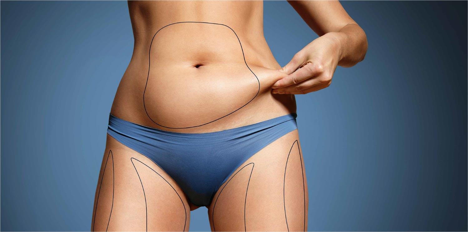
Abdominoplasty is a body-contouring procedure that involves much more than removing excess abdominal skin. The abdomen is made up of several interconnected layers, including skin, subcutaneous tissue, fascia and muscles. Each layer can contribute differently to the shape and appearance of the abdominal area.
For this reason, careful anatomical assessment is an important part of planning Abdominoplasty Dubai. Before developing an individual treatment approach, Dr. Shahram Sajjadi can assess the amount and distribution of loose skin, the underlying soft tissue, abdominal wall structure, muscle position, previous scars and the overall contour of the abdomen.
The assessment can also help distinguish between excess skin and changes involving the deeper abdominal wall. Recent literature describes abdominal contour concerns as involving separate superficial and deeper anatomical components, including skin laxity and changes in the musculoaponeurotic layer.
Understanding these differences allows treatment planning to be based on the patient's actual anatomy rather than simply the visible appearance of the abdomen.
Why Is Abdominal Anatomy Important Before Abdominoplasty?
The abdominal wall is a layered structure. It includes the skin and subcutaneous tissue on the surface, followed by fascia and the underlying abdominal muscles. The rectus abdominis muscles are positioned on either side of the midline and are enclosed by the rectus sheath. At the centre, connective tissues meet to form the linea alba.
Changes can occur at several of these levels. A patient may have loose skin after weight changes, while another may have stretching or separation involving the abdominal wall. Some patients may have a combination of these characteristics.
Therefore, the surgeon needs to determine which anatomical features are contributing to the patient's abdominal contour.
A detailed examination provides information that can influence the proposed treatment approach and helps establish realistic goals for the procedure.
How Is Abdominal Skin Assessed?
Skin assessment is one of the first parts of an abdominal examination. The surgeon looks at the amount, distribution and quality of excess skin rather than simply estimating how much tissue is present.
The abdomen is often assessed while the patient is standing because gravity makes areas of skin laxity and tissue redundancy easier to appreciate. The lower abdomen, around the navel and along the sides can be examined for differences in skin distribution.
The skin may also be gently pinched to evaluate its mobility and laxity. A pinch test is one of the recognised methods used during abdominoplasty planning to assess skin redundancy and help determine the amount of tissue that may potentially be removed.
This assessment is important because not every patient has the same pattern of excess skin. Some have greater laxity below the navel, while others have more widespread changes across the abdominal area.
What Is the Difference Between Loose Skin and Abdominal Muscle Changes?
Loose skin and changes in the deeper abdominal wall are separate anatomical issues.
Superficial skin laxity involves the skin and subcutaneous tissues. It can become more noticeable after substantial weight loss, pregnancy or changes in body composition.
Muscular or fascial changes occur deeper within the abdominal wall. One example is rectus diastasis, in which the linea alba between the paired rectus muscles becomes widened and attenuated.
These conditions can occur together, but they should not automatically be considered the same problem.
A patient can have considerable loose skin without significant rectus separation, while another may have changes in the abdominal wall with comparatively limited skin excess. Careful examination helps distinguish these findings.
How Are the Rectus Abdominal Muscles Evaluated?
The rectus abdominis muscles are important to abdominal contour because they form a major part of the anterior abdominal wall.
During an assessment, the patient may be examined while lying down and asked to contract the abdominal muscles. This can make the position and behaviour of the rectus muscles easier to evaluate.
The surgeon may palpate the midline and the borders of the rectus muscles to assess abdominal wall structure. Current surgical literature describes physical examination with the patient lying down and contracting the abdominal wall as a method that can help identify and measure rectus diastasis.The examination is not simply about measuring a gap. The overall shape and movement of the abdominal wall are also considered.
What Is Rectus Diastasis?
Rectus diastasis refers to widening and thinning of the linea alba between the two rectus abdominis muscles. It is different from a conventional hernia because the underlying anatomical mechanism involves attenuation and widening of the midline connective tissue rather than a discrete opening in the abdominal wall.Rectus diastasis can be associated with changes in abdominal contour, particularly when the abdominal wall is contracted.
However, the presence of a separation does not by itself provide a complete description of the abdominal wall. Research has shown that abdominal protrusion and wall laxity can occur with or without measurable rectus diastasis.For this reason, Dr. Shahram Sajjadi can consider the complete abdominal wall rather than relying on one measurement alone.
How Is the Midline and Linea Alba Assessed?
The linea alba is the connective tissue structure running along the midline of the abdomen. It connects the abdominal wall structures on either side and is closely associated with the rectus muscles.
During assessment, the surgeon may examine the central abdominal contour and palpate the midline while the patient contracts the abdominal muscles.
The width and condition of this area can provide information about the abdominal wall. In selected cases, the distance between the rectus muscles may also be evaluated at different points along the abdomen.
Research on rectus diastasis has used measurements around the umbilicus and at locations above and below it to understand the extent of separation.
This demonstrates why assessment should cover the abdominal wall rather than focusing on a single location.
Why Is the Abdomen Examined Both Standing and Lying Down?
Different positions provide different information.
When standing, gravity can make loose skin and tissue distribution more visible. The surgeon can assess how the abdominal contour changes naturally and identify areas where the skin hangs or folds.
When lying down, the abdominal wall becomes easier to examine directly. Muscle contraction can also make structural differences more apparent.
Recent surgical literature recommends assessing abdominal contour in both standing and supine positions, with muscle contraction used when appropriate to evaluate the abdominal wall.Using both positions provides a more complete picture of the patient's anatomy.
How Is Subcutaneous Fat Considered?
The tissue between the skin and abdominal wall can also contribute to abdominal contour. During assessment, the surgeon considers the amount and distribution of subcutaneous fat in addition to skin laxity.
This is important because abdominal fullness does not always result from loose skin or muscle changes. Subcutaneous tissue can contribute significantly to the overall shape of the abdomen.
The assessment therefore looks at the different layers individually before considering how they interact.
This layered approach can help determine whether the patient's primary concern relates to skin, soft tissue, abdominal wall structure or a combination of factors.
How Are Abdominal Scars Evaluated?
Previous abdominal surgery can influence treatment planning.
The surgeon may look for scars from procedures such as caesarean delivery, laparoscopic surgery, hernia repair or other abdominal operations. The location, length and direction of existing scars can provide important information about the abdominal tissues.
Previous operations may also have altered the anatomy of the abdominal wall or the position of relevant structures. Contemporary abdominoplasty literature recommends specifically assessing previous abdominal scars as part of preoperative planning.This information can be incorporated into the overall surgical design.
How Is the Navel Considered During Assessment?
The umbilicus is an important anatomical landmark in abdominoplasty.
Its position, shape and relationship to surrounding abdominal skin can be examined before surgery. When excess skin is removed, the relationship between the remaining abdominal tissue and the umbilicus must be considered carefully during treatment planning.
The surgeon therefore assesses the navel alongside the skin envelope and abdominal wall rather than treating it as an isolated feature.
Its position can also help the surgeon understand the proportions of the upper and lower abdomen.
How Are Hernias Considered During Abdominal Assessment?
A physical examination can also look for abdominal wall defects, including possible ventral or umbilical hernias.
A hernia represents a different anatomical issue from loose skin or rectus diastasis. Identifying a possible hernia before surgery is therefore important because it may influence the overall treatment plan.
Published surgical guidance recommends examining patients for ventral hernias during preoperative abdominoplasty assessment. Imaging may be considered when a hernia is suspected or when the diagnosis is uncertain.This illustrates why abdominal assessment involves more than evaluating the visible skin.
When Might Imaging Be Considered?
A physical examination is often sufficient for evaluating straightforward abdominal wall characteristics. However, imaging can provide additional information in selected situations.
For example, imaging may be considered if the surgeon suspects a hernia or needs further clarification about the abdominal wall anatomy.
Ultrasound has also been used in clinical research to measure rectus separation at defined points along the abdominal wall.
The decision to use imaging depends on the individual findings rather than being automatically necessary for every patient.
How Does Pregnancy Affect Abdominal Assessment?
Pregnancy can produce changes in the abdominal skin and deeper abdominal wall.
The skin may become stretched, while the abdominal wall can also undergo changes involving the linea alba and rectus muscles. These changes can contribute to differences in abdominal contour after pregnancy.
A patient's pregnancy history is therefore relevant during consultation. The surgeon can consider the location of skin laxity, abdominal wall characteristics and the overall distribution of excess tissue.
Importantly, the assessment remains individual because the extent and pattern of post-pregnancy changes vary between patients.
How Does Significant Weight Loss Affect Abdominal Anatomy?
Major weight loss can leave excess skin and changes in the distribution of subcutaneous tissue.
Patients who have lost substantial amounts of weight may have a different abdominal contour from someone whose changes are primarily related to pregnancy or natural ageing.
The surgeon can evaluate how much loose skin remains, where it is concentrated and how it relates to the underlying abdominal wall.
Current literature specifically identifies post-weight-loss abdominal changes as an important consideration in modern abdominoplasty planning.This assessment helps establish whether the treatment plan should focus primarily on the skin envelope or also address deeper structural characteristics.
How Is Abdominal Muscle Tone Considered?
Muscle tone and the condition of the abdominal wall can influence the appearance of the abdomen.
During examination, the surgeon may ask the patient to contract the abdominal muscles and observe the resulting contour. Palpation can provide additional information about muscle position and the underlying fascial layer.
The aim is not simply to determine whether the patient has strong or weak muscles. Instead, the surgeon assesses how the abdominal wall behaves and whether there is evidence of stretching or separation.
This information can then be considered alongside the amount of excess skin and soft tissue.
How Does Anatomy Influence the Abdominoplasty Plan?
Once the skin, soft tissue and abdominal wall have been assessed, these findings can be brought together to create an individual treatment plan.
For one patient, the main anatomical concern may be significant lower abdominal skin laxity. Another may have more widespread skin redundancy. A third may have abdominal wall changes that require particular attention during planning.
The procedure can therefore be tailored according to:
- The location and amount of excess skin
- Skin elasticity and mobility
- Subcutaneous tissue distribution
- Rectus muscle position
- Linea alba characteristics
- Abdominal wall contour
- Umbilical position
- Previous abdominal scars
- Possible hernias
- Overall body proportions
- The patient's individual goals
Modern approaches to abdominoplasty emphasise this layered anatomical evaluation rather than treating abdominal contour as a single problem.
Why Is Personalised Assessment Important?
There is no single abdominal anatomy shared by all patients. The amount of skin, distribution of tissue and condition of the abdominal wall can vary significantly.
A personalised assessment allows Dr. Shahram Sajjadi to understand these differences before discussing the appropriate surgical approach.
This is particularly important when patients compare themselves with photographs of other people. A similar-looking abdominal concern may have a completely different anatomical cause in another individual.
For someone researching the Best abdominoplasty Dubai, the quality of the consultation and anatomical assessment is therefore an important part of evaluating treatment options.
What Can Patients Discuss During the Consultation?
Patients can discuss their main concerns, previous pregnancies, weight changes, previous abdominal procedures and the type of abdominal contour they would like to achieve.
It can also be useful to explain whether the concern is primarily loose skin, abdominal fullness, changes around the navel, or a broader change in abdominal shape.
Dr. Shahram Sajjadi can then combine the patient's goals with clinical examination findings to determine which anatomical factors are most relevant to the treatment plan.
A consultation is therefore not simply a discussion about removing skin. It is an opportunity to understand how the different layers of the abdominal wall contribute to the patient's individual appearance.
Conclusion
Abdominal skin and muscle assessment is a fundamental part of planning Abdominoplasty Dubai. The abdomen consists of multiple anatomical layers, and changes in the skin, subcutaneous tissue, fascia and muscles can contribute differently to its appearance.
During assessment, Dr. Shahram Sajjadi can examine the abdomen in different positions, evaluate skin laxity, assess the underlying abdominal wall, consider the rectus muscles and linea alba, and review factors such as previous scars, pregnancy history, weight changes and possible hernias.
Understanding these individual anatomical characteristics allows the treatment plan to be based on the patient's actual structure rather than a standard approach. For patients considering Best abdominoplasty Dubai, a detailed consultation can help clarify how abdominal skin and muscle anatomy may influence their personalised treatment plan.
Frequently Asked Questions
What is assessed before abdominoplasty?
The assessment can include abdominal skin laxity, subcutaneous tissue, abdominal wall structure, rectus muscles, linea alba, umbilical position, previous scars and possible abdominal wall defects.
Why is abdominal skin assessed before surgery?
Skin assessment helps determine the location and extent of excess skin and how the skin envelope contributes to the patient's abdominal contour. A pinch test may also be used to assess skin laxity.
How are abdominal muscles examined?
The surgeon may examine the abdominal wall while the patient is lying down and may ask the patient to contract the abdominal muscles. Palpation can help identify the position of the rectus muscles and assess the midline.
What is rectus diastasis?
Rectus diastasis is widening and attenuation of the linea alba between the two rectus abdominis muscles. It can influence abdominal contour and is assessed as part of the deeper abdominal wall examination.
Can someone have loose abdominal skin without rectus diastasis?
Yes. Skin laxity and rectus diastasis are different anatomical conditions and can occur independently or together.
Is abdominal assessment performed while standing?
Yes. Standing assessment can make skin redundancy and the distribution of abdominal tissue easier to evaluate. The abdomen may also be assessed while lying down to examine the abdominal wall more closely.
Are previous abdominal scars considered?
Yes. Previous caesarean sections, abdominal operations and other scars can be relevant to treatment planning because they may affect the existing abdominal anatomy and tissue distribution.
Is imaging always needed before abdominoplasty?
No. Physical examination may provide sufficient information in straightforward cases. Imaging may be considered when there is uncertainty about the abdominal wall or a possible hernia.
Does pregnancy affect abdominal muscle assessment?
Pregnancy can affect both the abdominal skin and deeper abdominal wall structures. Pregnancy history can therefore be relevant when assessing skin laxity, rectus position and overall abdominal contour.
How does weight loss affect abdominoplasty planning?
Significant weight loss can leave excess abdominal skin and alter the distribution of soft tissue. The surgeon can assess these changes and determine how they relate to the underlying abdominal wall.
Why is the navel assessed during abdominoplasty planning?
The navel is an important abdominal landmark. Its position and relationship with the surrounding skin and abdominal wall are considered when developing an individual treatment plan.
How does abdominal anatomy influence the treatment plan?
The findings from the skin, soft-tissue and abdominal wall examination help the surgeon understand which anatomical factors are contributing to the patient's contour. This information can then be used to develop a personalised approach based on the patient's individual structure and goals.

 English
 Arabic
 French
 Portuguese
 Deutsch
 Turkish
 Dutch
Dutch
 Italiano
 Russian
 Romaian
 Portuguese (Brazil)
 Greek
Greek
 Bengali