


Demystifying Immunosuppression: A Systematic Workflow for Analyzing Regulatory T Cells (Tregs) in the Tumor Microenvironment
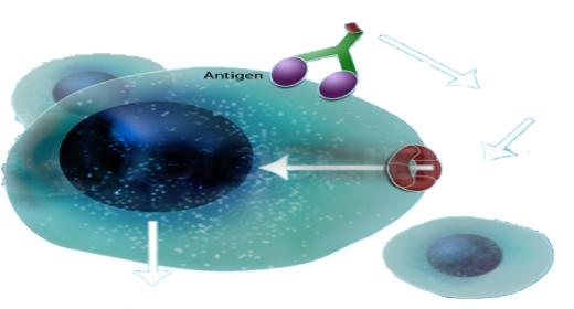
The advent of cancer immunotherapy has fundamentally shifted the paradigm of oncology. Treatments such as immune checkpoint inhibitors and CAR-T cell therapies have yielded unprecedented clinical responses. However, a significant portion of patients still experience primary or acquired resistance. Today, researchers agree that the major culprit behind this resistance is the highly complex, inherently immunosuppressive Tumor Microenvironment (TME).
At the center of this hostile environment are Regulatory T cells (Tregs). While physiologically essential for maintaining self-tolerance and preventing autoimmune diseases, Tregs are frequently hijacked by solid tumors. By infiltrating the TME, they suppress the activity of effector T cells (such as CD8+ cytotoxic T cells) and natural killer (NK) cells, effectively blinding the immune system to the malignancy. Consequently, targeting Tregs—either by depleting them or reprogramming their function—has become a top priority in biopharmaceutical R&D.
To successfully develop next-generation Treg-targeted therapies, researchers must adopt a systematic, multi-dimensional analytical workflow. Understanding these cells requires investigating three distinct biological pillars: their phenotypic identity, their suppressive function, and their spatial migration.
Step 1: Decoding Heterogeneity Through Phenotypic Analysis
The first major challenge in Treg research is cellular heterogeneity. Tregs are not a uniform population; they exist in various activation states and distinct subpopulations, each with unique molecular signatures. While classical Tregs are identified by the CD4+ CD25+ FoxP3+ phenotype, tumor-infiltrating Tregs often exhibit upregulated expression of specific immune checkpoints (such as CTLA-4, PD-1, TIGIT, and LAG-3) compared to their peripheral counterparts.
To accurately characterize these cells, researchers must establish a clear static phenotype. Conducting comprehensive Treg subpopulation expression profiling is a critical first step. By utilizing advanced multi-color flow cytometry, single-cell RNA sequencing (scRNA-seq), and multiplex immunohistochemistry, scientists can map the exact surface antigens and intracellular markers of Tregs within a specific tumor model. This phenotypic mapping is vital for identifying novel, tumor-specific Treg targets that allow for the selective depletion of malignant Tregs without triggering systemic autoimmunity.
Step 2: Evaluating Mechanisms of Immunosuppression
Identifying the presence of Tregs is only half the battle; understanding precisely how they neutralize anti-tumor immunity is the other. Tregs execute their suppressive mandate through a variety of contact-dependent and contact-independent mechanisms. These include the secretion of immunosuppressive cytokines (like TGF-β and IL-10), the consumption of vital survival cytokines (such as IL-2) to starve effector cells, and the direct induction of apoptosis in effector T cells via the perforin/granzyme pathway.
Because these mechanisms can vary widely depending on the tumor type, morphological identification must be followed by dynamic functional validation. Researchers utilize rigorous Treg functional profiling in tumors to measure the exact suppressive capacity of these cells in vitro and in vivo. These functional co-culture assays typically evaluate how effectively isolated Tregs can inhibit the proliferation and cytokine production of conventional CD4+ and CD8+ T cells. For drug developers, these assays are indispensable for validating whether a new immunotherapeutic compound can successfully block Treg-mediated suppression and restore effector T cell activity.
Step 3: Tracking Spatial Dynamics and Tumor Infiltration
A fundamental question in immuno-oncology is how Tregs accumulate in the TME in the first place. Tumors actively engineer their own protection by secreting specific chemokines (such as CCL22 and CCL28) that bind to corresponding receptors (like CCR4 and CCR10) on the surface of Tregs, heavily recruiting them from the peripheral blood into the tumor bed.
Interrupting this chemotactic recruitment represents a highly promising therapeutic strategy. To screen drug candidates that block this pathway, scientists rely on specialized Tregs migration assays. These transwell and microfluidic-based chemotaxis assays allow researchers to quantify the movement of Tregs toward tumor-derived chemokine gradients in real-time. By understanding and successfully inhibiting this migratory behavior, drug developers can theoretically prevent the immunosuppressive shield from forming around the tumor.
Bridging Preclinical Research to Clinical Success
Moving a Treg-targeted therapy from the bench to the bedside requires robust preclinical data. The triad of evaluating expression profiles, functional mechanisms, and migratory behaviors creates a scientifically rigorous, closed-loop research workflow.
Executing these sophisticated assays requires advanced laboratory infrastructure and highly specialized immunological expertise. Many biopharmaceutical companies and academic institutions opt to collaborate with specialized Contract Research Organizations (CROs) to accelerate this process. Industry partners like Creative Biolabs provide researchers with access to these optimized immune-oncology platforms, ensuring that the data driving early-stage drug discovery is both highly reproducible and biologically relevant.
As our understanding of the TME deepens, systematically unraveling the biology of Regulatory T cells will remain critical. By meticulously profiling who these cells are, what they do, and how they travel, the scientific community moves one step closer to dismantling the immune barriers of cancer.

 English
 Arabic
 French
 Spanish
 Portuguese
 Deutsch
 Turkish
 Dutch
Dutch
 Italiano
 Romaian
 Portuguese (Brazil)
 Greek
Greek
 Bengali