


Lipoma Treatment in Riyadh: The Value of Diagnostic Imaging
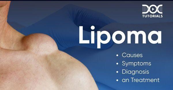
Lipoma Treatment in Riyadh: The Value of Diagnostic Imaging
Lipoma Treatment in Riyadh integrates advanced diagnostic imaging as a cornerstone of clinical practice, ensuring that every patient receives a tailored and precise treatment plan. While many lipomas can be identified through a straightforward physical examination due to their characteristic "doughy" or "rubbery" texture and mobility, the medical standard in the region prioritizes objectivity. By utilizing sophisticated imaging technologies, clinicians can confirm the diagnosis, map the anatomical relationships of the growth, and plan the surgical approach with a high degree of confidence, ultimately enhancing safety and procedural success.
The Role of Ultrasound in Initial Assessment
Ultrasound is frequently the first-line diagnostic tool employed for subcutaneous masses. As a non-invasive, radiation-free, and highly accessible technology, it allows the clinician to visualize the lipoma in real-time. Through ultrasound, the provider can differentiate a lipoma from other conditions such as sebaceous cysts, hernias, or other soft-tissue anomalies. The imaging reveals the mass’s echogenicity—a measurement of how it reflects sound waves—which helps confirm the presence of fatty tissue. Furthermore, it allows the clinician to define the mass's boundaries, assess its depth relative to the skin, and determine its proximity to critical structures like underlying fascia or superficial nerves.
Advanced Imaging for Complex Cases
In instances where a lipoma presents with atypical features—such as being larger than a few centimeters, located in a challenging anatomical area like the neck or axilla, or feeling firm or fixed to deeper tissues—advanced imaging modalities become essential. Magnetic Resonance Imaging (MRI) is considered the gold standard for soft-tissue assessment. It provides high-resolution, multi-planar views that map the lipoma’s relationship to adjacent muscles, neurovascular bundles, and other deep-seated structures.
Computed Tomography (CT) scans may also be utilized, particularly when there is a need to rule out deeper extensions or when an MRI is not suitable for the patient. These advanced tools offer a comprehensive "blueprint" for the surgeon. By understanding the exact location and extent of the lipoma before the first incision is made, the surgical team can minimize the duration of the procedure, reduce tissue trauma, and ensure that the entire capsule of the lipoma is addressed, which is critical for preventing recurrence.
Clinical Decision-Making
Diagnostic imaging does more than just confirm the presence of a lipoma; it informs the entire clinical strategy. For example, imaging might reveal that a lipoma is "intermuscular," meaning it grows between muscle fibers. Knowing this allows the surgeon to prepare for a more specialized dissection rather than a simple surface excision. Conversely, if imaging confirms that a mass is superficial and well-encapsulated, the clinician can confidently proceed with a minimally invasive, smaller-incision approach, which is ideal for achieving superior aesthetic outcomes with minimal scarring.
Prioritizing Patient Safety
The commitment to diagnostic precision serves as a vital safeguard for the patient. It ensures that the procedure is always clinically indicated and performed with full knowledge of the local anatomy. By removing the guesswork, medical professionals in Riyadh protect patients from unnecessary complications. Furthermore, this rigorous approach is instrumental in the rare, proactive effort to identify "red flags" that might suggest a more concerning condition, such as a liposarcoma. Although benign lipomas are the norm, diagnostic imaging provides the evidence-based confidence that the treatment plan is correct, shielding the patient from diagnostic ambiguity.
Bridging the Gap to Successful Outcomes
The integration of diagnostic imaging into the standard of care reflects a broader commitment to excellence in Riyadh’s medical sector. It empowers the patient with knowledge, as the medical team can show them the findings and explain the rationale for the selected surgical technique. This transparency fosters trust and aligns expectations between the patient and the provider. By investing in these diagnostic steps, the medical community ensures that Lipoma Treatment in Riyadh is not just an effective procedure, but a precise, safe, and highly predictable experience that prioritizes the patient’s long-term health and wellness. Ultimately, diagnostic imaging turns a routine concern into a well-managed, successful medical intervention, providing the clarity and assurance that every patient deserves.

 English
 Arabic
 French
 Spanish
 Portuguese
 Deutsch
 Turkish
 Dutch
Dutch
 Italiano
 Romaian
 Portuguese (Brazil)
 Greek
Greek
 Bengali